


雷酚内酯 促销 ≥98%(HPLC),分析标准品
促销价¥{{model.attbuying.price}}
市场价¥0.00
累计销量0 累计评价0 人评论
-
{{item.title}}
| 包装规格: | 20mg in glass bottle |
| 产品简介: | 一种具有口服活性的雄激素受体 (AR) 的泛拮抗剂,对人野生型 AR 的 IC50 为 467 nM。Triptophenolide 可降低 AR 表达、抑制 AR 核转位、下调前列腺特异性抗原 mRNA 水平,并抑制 AR 阳性前列腺癌细胞的增殖。Triptophenolide 对乳腺癌具有抗肿瘤作用,其机制包括抑制细胞增殖和迁移,诱导G1期阻滞和细胞凋亡,以及抑制异种移植瘤的生长。Triptophenolide 可抑制细胞焦亡、减轻组织炎症并改善滑膜损伤。Triptophenolide 可用于前列腺癌、类风湿性关节炎和乳腺癌的研究。 |
| 溶解性: | DMSO:100 mg/mL (320.10 mM;超声助溶) |
| 储备液保存: | -80°C, 2 years; -20°C, 1 year。 |
| 体内实验: | 1、请依序添加每种溶剂: 10% DMSO→90% Corn Oil Solubility: ≥ 2.5 mg/mL (8.00 mM); 澄清溶液 此方案可获得 ≥ 2.5 mg/mL(饱和度未知)的澄清溶液,此方案实验周期在半个月以上的动物实验酌情使用。 以 1 mL 工作液为例,取 100 μL 25.0 mg/mL 的澄清 DMSO 储备液加到 900 μL玉米油中,混合均匀。 注:工作液建议您现用现配,当天使用。 <1mg/ml表示微溶或不溶。 普西唐提供的所有化合物浓度为内部测试所得,实际溶液度可能与公布值有所偏差,属于正常的批间细微差异现象。 请根据产品在不同溶剂中的溶解度选择合适的溶剂配制储备液;一旦配成溶液,请分装保存,避免反复冻融造成的产品失效。 |
| 靶点: | Caspase 3;Caspase-9;Bax;Bak;Bim |
| 体外研究: | Triptophenolide (50 nM-5 μM) 可强效抑制 PC-3 细胞中 DHT 诱导的野生型 AR、AR F876L、AR T877A 及 W741C+T877A AR 双突变体的转录活性,其 IC50 值分别为 260 nM、480 nM、388 nM 和 437 nM。在无 DHT 的条件下,该化合物对这些 AR 变体无激动活性。 Triptophenolide (500 nM-5 μM) 可显著下调 LNCaP 细胞中前列腺特异性抗原 mRNA 的表达。 Triptophenolide (0.1-50.0 μM) 可剂量依赖性地抑制 AR 阳性 LNCaP 前列腺癌细胞的生长,而对 AR 阴性 PC-3 前列腺癌细胞的生长无显著影响。 Triptophenolide (10 nM-10 μM) 可在无细胞体系中竞争性结合雄激素受体配体结合域,其 IC50 为 467 nM。 Triptophenolide (50 nM-5 μM; 24 h) 可剂量依赖性地抑制 LNCaP 细胞中雄激素受体蛋白的表达,处理 24 小时后,在 500 nM 和 5 μM 浓度下作用显著。 Triptophenolide (5 μM) 可有效抑制 LNCaP 细胞中 DHT 诱导的雄激素受体核转位。 Triptophenolide (0-400 μg/mL;24/48 h) 可浓度和时间依赖性地抑制 MCF-7 和 MDA-MB-231 乳腺癌细胞的增殖。 Triptophenolide (MCF-7 细胞为 180.3 μg/mL、MDA-MB-231 细胞为 322.5 μg/mL;48 h) 可显著下调检测到的靶基因 mRNA 水平,并降低 MCF-7 和 MDA-MB-231 乳腺癌细胞中相应的蛋白表达。 Triptophenolide (150 μg/mL;24 h/48 h) 可诱导 MCF-7 和 MDA-MB-231 乳腺癌细胞发生凋亡及 G1 期细胞周期阻滞,并显著抑制细胞迁移。 |
| 体内研究: | Triptophenolide (5-15 mg/kg;灌胃;每日 1 次;连续 15 天) 可显著降低胶原抗体诱导的类风湿性关节炎小鼠的关节炎评分,抑制滑膜炎症因子水平,并减轻滑膜损伤。 Triptophenolide (10 mg/kg;腹腔注射;每3天一次;共21天) 显著抑制 MCF-7 异种移植瘤的生长,减轻肿瘤重量,并延长荷瘤 BALB/c 裸鼠的生存期。 |
| 细胞实验: | Western Blot Analysis Cell Line:LNCaP cells Concentration:50 nM, 500 nM, 5 μM Incubation Time:24 h Result:Effectively suppressed AR protein expression in a dose-dependent manner. Showed significant suppression at 500 nM and 5 μM. RT-PCR Cell Line:MCF-7 and MDA-MB-231 breast cancer cells Concentration: 180.3 μg/mL for MCF-7 cells and 322.5 μg/mL for MDA-MB-231 cells Incubation Time:48 h Result:significantly upregulates the mRNA expression of multiple pro-apoptotic genes (e.g., BIM, BAK1, BAX, CASP3/9, TP53) and downregulates proliferation-associated genes such as AKT1 in both MCF-7 and MDA-MB-231 breast cancer cells. Cell Viability Assay Cell Line:MCF-7 and MDA-MB-231 breast cancer cells Concentration:0, 100, 200, 300, 400 μg/mL Incubation Time:24/48 h Result:Inhibits proliferation in MCF-7 and MDA-MB-231 breast cancer cells Western Blot Analysis Cell Line:MCF-7 and MDA-MB-231 breast cancer cells Concentration:180.3 μg/mL for MCF-7 cells and 322.5 μg/mL for MDA-MB-231 cells Incubation Time:48 h Result:significantly upregulated the expression of pro-apoptotic proteins BIM, BAK1, BAX, and CYCS in both MCF-7 and MDA-MB-231 breast cancer cells. Cell Cycle Analysis Cell Line:MCF-7 and MDA-MB-231 breast cancer cells Concentration:150 μg/mL Incubation Time:24 h/48 h Result:Induced G1-phase cell cycle arrest in both MCF-7 and MDA-MB-231 breast cancer cells, with a marked increase in G1-phase cell proportion and a corresponding decrease in S-phase cell proportion. Apoptosis Analysis Cell Line:MCF-7 and MDA-MB-231 breast cancer cells Concentration:150 μg/mL Incubation Time:24 h/48 h Result:Significantly induced apoptosis in both MCF-7 and MDA-MB-231 breast cancer cells, with markedly elevated total apoptosis rates. Cell Proliferation Assay Cell Line:MCF-7 and MDA-MB-231 breast cancer cells Concentration:150 μg/mL Incubation Time:24 h/48 h Result:Significantly and time-dependently inhibited the migration of both MCF-7 and MDA-MB-231 breast cancer cells, with markedly reduced migration rates at 12 h and 24 h. |
| 动物实验: | Animal Model:BALB/c nude mice (8-10-week-old) Dosage:10 mg/kg Administration:intraperitoneal injection; every 3 days; 21 days Result:Significantly inhibited MCF-7 xenograft tumor growth, reduced tumor volume and weight, and prolonged the survival of tumor-bearing mice compared to the DMSO control group, with statistically significant improvements. Animal Model:BALB/c (8-10-week-old) Dosage:5 mg/kg; 15 mg/kg Administration:intragastric; once daily; 15 days Result:Significantly reduced mouse arthritis scores compared to the RA group at matching time points. Suppressed synovial tissue expression of inflammatory factors IL-1β, IL-6, and TNF-α, with levels lower than those in the RA group. Alleviated synovial injury and reduced inflammatory lesions in joint tissue, with statistically significant improvements compared to the RA group. |
| 保存条件: | 2-8℃ |
-
蜀牛玻璃三角瓶
¥85.00 销量:16
-
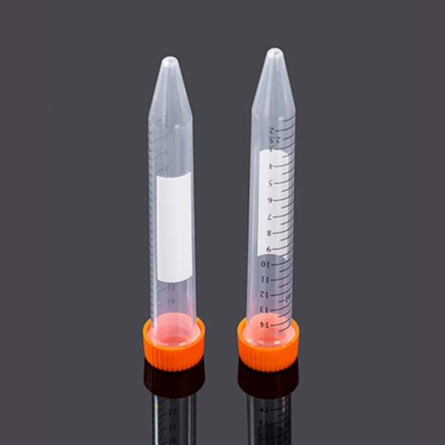 15mL锥底离心管,袋装灭菌,橙色盖
¥636.00 销量:12
-
UFC5030BK 超滤管[0.5ml 30KD]
¥52.00 销量:10
-
UFC5010BK 超滤管[0.5ml 10KD]
¥52.00 销量:10
-
20×TBST缓冲液
¥130.00 销量:10
-
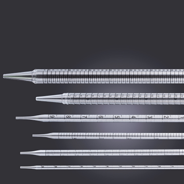 10mL移液管,易撕型全塑包装
¥456.00 销量:10
-
¥158.00
通用型 DNA 纯化回收试剂盒 -
¥158.00
质粒快速小提试剂盒 -
¥828.00
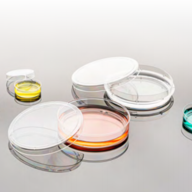
100mm TC细胞培养皿,袋装灭菌