

异夏佛塔苷 ≥98%(HPLC),分析标准品
促销价¥{{model.attbuying.price}}
市场价¥0.00
累计销量0 累计评价0 人评论
别称
{{model.alias}}
中文名称
{{model.name}}
英文名称
{{model.enname}}
品牌
{{model.brand}}
CAS
{{model.cas}}
分子式
分子量
{{model.molecularweight}}
MDL
{{model.mdl}}
货号
{{model.procode}}
-
{{item.title}}
看了又看
| 包装规格: | 10mg in glass bottle |
| 产品简介: | 是从 Desmodium uncinatum 根部分泌物中发现的 C-糖基黄酮类化合物,能抑制 S. hermonthica 胚芽的生长。Isoschaftoside 能够降低衰老细胞活性氧 (ROS),并诱导细胞增殖。Isoschaftoside 激活自噬 (autophagy)。Isoschaftoside 可用于抗肿瘤、抗炎、抗氧化、抗高血压、保肝和杀线虫等的研究。 |
| 溶解性: | DMSO : 100 mg/mL (177.15 mM; 超声助溶) |
| 储备液保存: | -80°C, 6 months -20°C, 1 month (protect from light) |
| 体内实验: | 1、请依序添加每种溶剂: 10% DMSO→40% PEG300→5% Tween-80→45% Saline Solubility: ≥ 2.5 mg/mL (4.43 mM); 澄清溶液 此方案可获得 ≥ 2.5 mg/mL(饱和度未知)的澄清溶液。 以 1 mL 工作液为例,取 100 μL 25.0 mg/mL 的澄清 DMSO 储备液加到 400 μL PEG300 中,混合均匀;再向上述体系中加入 50 μL Tween-80,混合均匀;然后再继续加入 450 μL 生理盐水 定容至 1 mL。 2、请依序添加每种溶剂: 10% DMSO→90% (20% SBE-β-CD in Saline) Solubility: ≥ 2.5 mg/mL (4.43 mM); 澄清溶液 此方案可获得 ≥ 2.5 mg/mL(饱和度未知)的澄清溶液。 以 1 mL 工作液为例,取 100 μL 25.0 mg/mL 的澄清 DMSO 储备液加到 900 μL 20% 的 SBE-β-CD 生理盐水水溶液 中,混合均匀。 2 g SBE-β-CD(磺丁基醚 β-环糊精)粉末定容于 10 mL 的生理盐水中,完全溶解至澄清透明。 <1mg/ml表示微溶或不溶。 普西唐提供的所有化合物浓度为内部测试所得,实际溶液度可能与公布值有所偏差,属于正常的批间细微差异现象。 请根据产品在不同溶剂中的溶解度选择合适的溶剂配制储备液;一旦配成溶液,请分装保存,避免反复冻融造成的产品失效。 |
| 体外研究: | Isoschaftoside (4 μM,12 天) 通过降低衰老成纤维细胞中 RAC2 和 LINC00294 的表达来抑制活性氧 (ROS)。 Isoschaftoside (0.25-4 μM,12 天) 显著增加衰老成纤维细胞的细胞增殖。 Isoschaftoside (1 μM,12 天) 显著提高衰老成纤维细胞线粒体膜电位 (MMP),诱导代谢变化,恢复线粒体功能,并降低细胞对糖酵解的依赖。 Isoschaftoside (1 μM,12 天) 减少衰老成纤维细胞 DNA 损伤和碎片化,激活自噬系统,降低细胞中炎症因子的表达。 Isoschaftoside (20-600 μg/mL) 对M. incognita具有强效的杀线虫活性,(LC50 = 323.09 μg/mL)。 Isoschaftoside (1-500 μM,24 小时) 在 Palmitic acid (PA) 诱导的 HepG2 自噬抑制中通过激活自噬通量减少脂质沉积,抑制轻链 3-II (LC3-II) 和 SQSTM1/p62 的表达。 Isoschaftoside (0.05-2 mg/mL) 显著降低猪肾近端小管基底外侧膜的 Na+、K+-ATPase 活性。 Isoschaftoside (0-1000 μM,9-24 小时) 可抑制 Lipopolysaccharides (LPS) 激活的小胶质细胞炎症反应。 |
| 体内研究: | Isoschaftoside (20 mg/kg;腹腔注射;每日一次,持续 4 周) 可逆转小鼠的非酒精性脂肪性肝病 (NAFLD) 并减少肝脏脂肪变性。 |
| 细胞实验: | Cell Proliferation Assay Cell Line: human dermal fibroblasts Concentration: 0.25, 0.5, 1, 2, 4 μM Incubation Time: 12 days Result: The increase in cell proliferation rate was most significant at a concentration of 1 μM. Cell Autophagy Assay Cell Line: human dermal fibroblasts Concentration: 1 μM Incubation Time: 12 days Result: LC3B was noticeably activated. Perinuclear mitochondria were reduced. Showed significantly enhanced levels of autophagy. Significant reduction in lipofuscin was observed. Western Blot Analysis Cell Line: HepG2 cells Concentration: 1, 10, 20, 50, 100, 200, 300, 400, 500 μM Incubation Time: 24 h Result: When the concentration was higher than 200 μM, observed significant reductions in LC3-II expression. Decreased LC3-II and p62 expression in Palmitic acid (PA)-induced autophagy. Western Blot Analysis Cell Line: BV2 microglial cells Concentration: 200 μM Incubation Time: 9 h or 24 h Result: Significantly inhibited Lipopolysaccharides-induced iNOS, TNF-α, IL-1β, and COX2 protein expression. Significantly reduced the Lipopolysaccharides-induced HIF-1α, HK2, and PFKFB3 expression. Attenuated Lipopolysaccharides-induced phosphorylation of ERK1/2 and mTOR. |
| 动物实验: | Animal Model: C57 mice fed with a high-fat diet (HFD) (4 weeks; male; C57BL/6JGpt) Dosage: 20 mg/kg Administration: Intraperitoneal injection (i.p.); daily for 4 weeks Result: Significantly reduced the body weight and body size of the high-fat diet mice compared with the model group. Reduced the visceral fat weight of high-fat diet mice. Significantly attenuated hepatic lipid accumulation induced by a high-fat diet and decreased the number of intracellular lipid droplets and hepatocyte balloons. Reversed the damage to liver function and the elevation of blood glucose caused by a high-fat diet. The expression level of p62 and LC3-II was significantly lower than that in the HFD group. |
| 保存条件: | 2-8℃ |
| 注意事项: | 1、为了您的安全和健康,请穿实验服并戴一次性手套操作。 2、以上信息仅做参考交流之用。 |
-
蜀牛玻璃三角瓶
¥85.00 销量:16
-
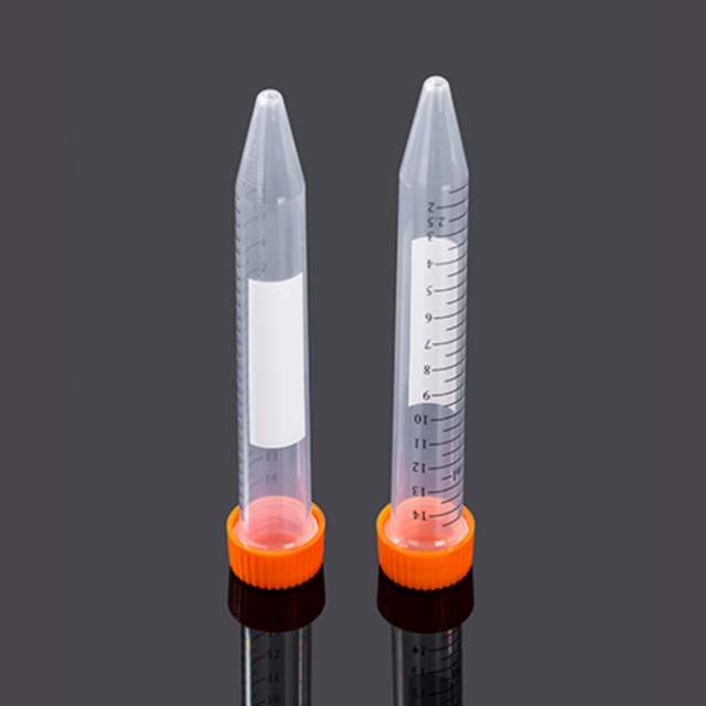 15mL锥底离心管,袋装灭菌,橙色盖
¥636.00 销量:12
-
UFC5030BK 超滤管[0.5ml 30KD]
¥52.00 销量:10
-
UFC5010BK 超滤管[0.5ml 10KD]
¥52.00 销量:10
-
20×TBST缓冲液
¥130.00 销量:10
-
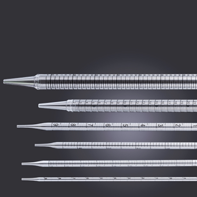 10mL移液管,易撕型全塑包装
¥456.00 销量:10
猜你喜欢
-
¥158.00
通用型 DNA 纯化回收试剂盒 -
¥158.00
质粒快速小提试剂盒 -
¥828.00
100mm TC细胞培养皿,袋装灭菌