

Saracatinib 促销 99.5778%
促销价¥{{model.attbuying.price}}
市场价¥0.00
累计销量0 累计评价0 人评论
-
{{item.title}}
| 包装规格: | 5mg 25mg 100mg in glass bottle |
| 溶解性: | 溶于DMSO(28 mg/mL 超声) |
| 产品描述: | 基本信息 产品编号:P10805 产品名称:PP1 CAS: 172889-26-8 储存条件 粉末 -20℃ 四年 分子式: C16H19N5 溶于液体 -80℃ 六个月 分子量 281.36 -20℃ 一个月 化学名: Solubility (25°C) 体外 DMSO 5mg/mL (17.77mM) Ethanol Insoluble Water Insoluble 体内(现配现用) <1mg/ml表示微溶或不溶。 普西唐提供的所有化合物浓度为内部测试所得,实际溶液度可能与公布值有所偏差,属于正常的批间细微差异现象。 请根据产品在不同溶剂中的溶解度选择合适的溶剂配制储备液;⼀旦配成溶液,请分装保存,避免反复冻融造成的产品失效。 制备储备液 浓度 溶液体积 质量 1mg 5mg 10mg 1mM 3.5542mL 17.7708mL 35.5417mL 5mM 0.7108mL 3.5542mL 7.1083mL 10mM 0.3554mL 1.7771mL 3.5542mL 生物活性 产品描述 一种有效的选择性 Src 家族抑制剂,抑制 Lck 和 Fyn,IC50 分别为 5 和 6nM。 靶点/IC50 IC50: 5nM (Lck), 6nM (Fyn), 250 nM (EGFR), >50μM (JAK2 体外研究 PP1 inhibits Lck (IC50=5nM) and FynT (IC50=6nM) in vitro at concentrations significantly lower than those required to inhibit ZAP-70 (IC50>100μM), JAK2 (IC50>50μM), the EGFR kinase, and protein kinase A. PP1 inhibits whole cell tyrosine phosphorylation and proliferation in T cells stimulated with anti-CD3 and mitogens. PP1 selectively inhibits IL-2 gene expression over GM-CSF and IL-2R gene induction in human T cells. 推荐实验方法(仅供参考) 激酶实验: Protein A-Sepharose beads (prepared as a 50% (w/v) suspension) are added to the antibody/lysate mixture at 250μL/mL and allowed to incubate for 30 min at 4°C. The beads are then washed twice in 1mL of lysis buffer and twice in 1 mL of kinase buffer (25 mM HEPES, 3mM MnCl2, 5mM MgCl2, and 100μM sodium orthovanadate) and resuspended to 50% (w/v) in kinase buffer. Twenty-five microliters of the bead suspension is added to each well of the enolase-coated 96-well high protein binding plate together with an appropriate concentration of compound and [γ-32P]ATP (25μL/well of a 200μCi/mL solution in kinase buffer). After incubation for 20 min at 20°C, 60μL of boiling 2× solubilization buffer containing 10mM ATP is added to the assay wells to terminate the reactions. Thirty microliters of the samples is removed from the wells, boiled for 5 min, and run on a 7.5% SDS-polyacrylamide gel. The gels are subsequently dried and exposed to Kodak X-AR film. For quantitation, films are scanned using a Molecular Dynamics laser scanner, and the optical density of the major substrate band, enolase p46, is determined. Concentrations of compound that causes 50% inhibition of enolase phosphorylation (IC50) are determined from a plot of the density versus concentration of compound. In companion experiments for measuring the activity of compounds against Lck, the assay plate is washed with two wash cycles on a Skatron harvester using 50mM EDTA, 1mM ATP. Scintillation fluid (100μL) is then added to the wells,and P incorporation is measured using a Pharmacia Biotech micro-β-counter. Concentrations of compound that causes 50% inhibition of enzyme activity (IC50) are determined from a plot of the percent inhibition of enzyme activity versus concentration of compound. 细胞实验: Inhibition of anti-CD3-stimulated tyrosine phosphorylation in purified human peripheral blood T cells is measured as follows. All incubations are carried out at 37°C in an Eppendorf Thermomixer 5436 at a mixing setting of 11. Cells (1×10⁶ in 100 μL of RPMI 1640 medium) are incubated for 15 min with drug prior to a 6-min incubation with 1 μg of anti-CD3/mL (antileu4, 100μg/mL). The final volume of the reaction is 115μL.Reactions are terminated by the addition of 57.5μL of 3×solubilization buffer incubated at 100°C prior to its addition. Samples are mixed, boiled for 5 min, and stored at -70°C. Western blots of these cell lysates, run on 10% SDS-polyacrylamide gels, are probed with a polyclonal anti-phosphotyrosine antibody, and immune complexes are detected with I-labeled protein A (ICN). For quantitation, films are scanned using a Molecular Dynamics laser scanner, and the optical densities of the major substrate band, p70,are quantitated in the presence of anti-CD3 (in the presence and absence of drug).Percent inhibition is calculated as follows: (1-(p70 optical density units in presence of drug/p70 units in absence of drug))×100.IC50 equals the concentration of compound at which 50% inhibition is measured. |
| 保存条件: | -20℃ |
-
蜀牛玻璃三角瓶
¥85.00 销量:16
-
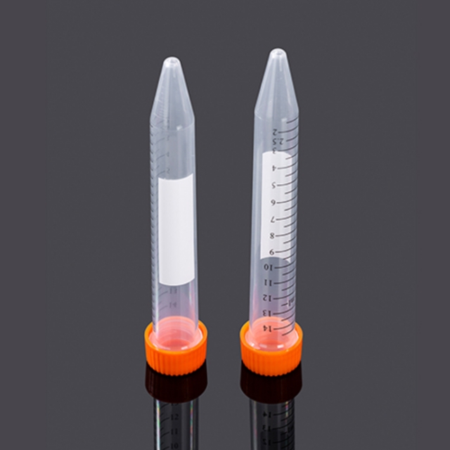 15mL锥底离心管,袋装灭菌,橙色盖
¥636.00 销量:12
-
UFC5030BK 超滤管[0.5ml 30KD]
¥52.00 销量:10
-
UFC5010BK 超滤管[0.5ml 10KD]
¥52.00 销量:10
-
20×TBST缓冲液
¥130.00 销量:10
-
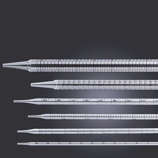 10mL移液管,易撕型全塑包装
¥456.00 销量:10


-
¥158.00
通用型 DNA 纯化回收试剂盒 -
¥158.00
质粒快速小提试剂盒 -
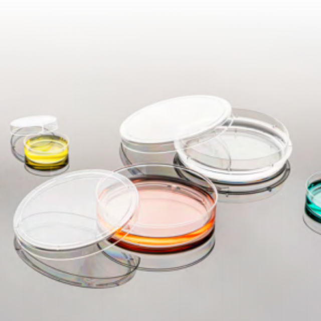
¥828.00
100mm TC细胞培养皿,袋装灭菌